Sunday, January 18, 2015

Leukocytes Agranulocytes Monocytes


Blood smears


Monocyte - Greater increase

Leukocyte Agranulocyte Monocyte

* It is presented as the greatest of leukocytes.
* Absence of granules, bluish cytoplasm.
* Core reniform (kidney-shaped).

Black Circle - Monocyte
Black Arrow - Erythrocyte

Red Arrow - Platelets


Leukocytes Agranulocytes Lymphocytes


Blood smears


Lymphocyte - Greater increase

Leukocyte Agranulocyte Lymphocyte

*These are usually classified as small, medium or large. Medium and large lymphocytes are generally seen mainly in fibrous connective tissue and only occasionally in the circulation bloodstream

Black Circle - Lymphocyte
Black Arrow - Erythrocyte
Red Arrow - Platelets


Lymphocyte - Greater increase

Leukocyte Agranulocyte Lymphocyte

*These are usually classified as small, medium or large. Medium and large lymphocytes are generally seen mainly in fibrous connective tissue and only occasionally in the circulation bloodstream

Black Circle - Lymphocyte
Black Arrow - Erythrocyte

Red Arrow - Platelets


Leukocytes Granulocytes Basophils


Blood smears

Basophil - Greater increase

Leukocyte Granulocyte Basophil

*They have a pale nucleus that is usually hidden by granules.

Black Circle - Basophil     
Black Arrow - Erythrocyte

Red Arrow - Platelets


Wednesday, January 14, 2015

Leukocytes Granulocytes Eosinophils


Blood smears


Eosinophil - Greater increase

Leukocyte Granulocyte Eosinophil

*These have large granules
*A prominent nucleus that is divided into two lobes
*Orange cytoplasm

Black Circle - Eosinophil  
Black Arrow - Erythrocyte
Red Arrow - Platelets


Eosinophil - Greater increase

Leukocyte Granulocyte Eosinophil

*These have large granules
*A prominent nucleus that is divided into two lobes
*Orange cytoplasm

Black Circle - Eosinophil  
Black Arrow - Erythrocyte
Red Arrow - Platelets


Leukocytes Granulocytes Neutrophils


Blood smears

Neutrophils - Greater increase

Leukocytes Granulocytes Neutrophils

* fine cytoplasmic granules
*variety of nuclear shapes

Black Circle - Neutrophils
Black Arrow - Erythrocyte


Neutrophils - Greater increase

Leukocytes Granulocytes Neutrophils

* fine cytoplasmic granules
*variety of nuclear shapes

Black Circle - Neutrophils
Black Arrow - Erythrocyte
Red Arrow - Platelets


 Neutrophils - Greater increase


Leukocytes Granulocytes Neutrophils

* fine cytoplasmic granules
*variety of nuclear shapes

Black Circle - Neutrophils
Black Arrow - Erythrocyte
Red Arrow - Platelets


Neutrophils stick - Greater increase

Leukocytes Granulocytes Neutrophils stick

* fine cytoplasmic granules
*not segmented core

Black Circle - Neutrophils stick
Black Arrow - Erythrocyte

Red Arrow - Platelets


Platelets


Blood smears


Platelets 100x

Platelets are disc-shaped cells circulating in the blood. They are involved in the cellular mechanisms of primary haemostasis leading to the formation of blood clots.

The normal number of platelets in the blood is 150,000 - 400,000 platelets per microliter (mcL). Normal value ranges may vary slightly. Some lab use different measurements or may test different specimens. Talk to your doctor about your test results.

Red Circles: Platelets

Black Arrows: Erythrocyte


Erythrocytes


Blood smears


Erythrocytes/ Red Blood Cells (RBCs) - 100x

An erythrocyte is a disc-shaped cell with a thick rim and a thin sunken centre and has two main functions:

1- To pick up oxygen from the lungs and deliver it to tissues elsewhere
2- To pick up carbon dioxide from other tissues and unload it in the lungs

Normal RBC range is:

Male: 4.7 to 6.1 million cells per microliter (cells/mcL)
Female: 4.2 to 5.4 million cells/mcL


Arrow - Platelets


Nerve - Slide 84


Nerve Tissue - Periphecal Nervous System

Special Color for Nerves: Masson's trichome


Nerve - Middle increase

Nerve - Nerve Tissue - Periphecal Nervous System - with Epineurium, Perineurium and Endoneurium.

- Fibers in Transversal Section
- Myelinated and Unmyelinated Nerve Fibers

1-Epineurium
2-Perineurium
3-Endoneurium


Nerve - Middle increase

Nerve - Nerve Tissue - Periphecal Nervous System - with Epineurium, Perineurium and Endoneurium.

- Fibers in Transversal Section
- Myelinated and Unmyelinated Nerve Fibers

1-Epineurium
2-Perineurium
3-Endoneurium
Black circle - Vasa Nervorum (blood vessel).


Nerve - Middle increase

Nerve - Nerve Tissue - Periphecal Nervous System - with Epineurium, Perineurium and Endoneurium.

- Fibers in Transversal Section
- Myelinated and Unmyelinated Nerve Fibers

1-Epineurium
2-Perineurium
3-Endoneurium
Black circle - Vasa Nervorum (blood vessel).


Nerve - Longitudinal Section - Middle increase

Nerve - Nerve Tissue - Periphecal Nervous System - with Epineurium, Perineurium and Endoneurium.

- Fibers in Longitudinal Section
- Myelinated and Unmyelinated Nerve Fibers


Connective Tissue is in blue and the Nerver in red.


Nerve - Slide 83


Nerve Tissue - Periphecal Nervous System


Nerve - Smaller increase

Nerve - Nerve Tissue - Periphecal Nervous System - with Epineurium, Perineurium and Endoneurium.
- Fibers in Transversal Section
- Myelinated and Unmyelinated Nerve Fibers

It shows Vasa Nervorum (blood vessel).


Nerve - Middle increase

Nerve - Nerve Tissue - Periphecal Nervous System - with Epineurium, Perineurium and Endoneurium.

- Fibers in Transversal Section
- Myelinated and Unmyelinated Nerve Fibers

1-Epineurium
2-Perineurium
3-Endoneurium

Black circle - Vasa Nervorum (blood vessel).


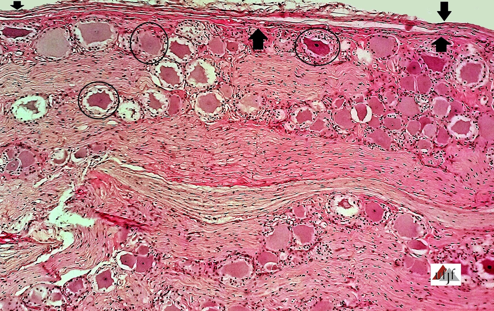
Ganglion - Slide 76


Nerve Tissue - Peripheral Nervous System


 Ganglion - Smaller increase


Ganglion - Nerve Tissue - Peripheral Nervous System with:

- Modeled Dense Connective Itself Tissue Fibrous/Capsular (arrow)
- Neurons Bodies (Black Circle)
- Satellite cells (they are small around Neurons)
- Nerve Fibers.


Ganglion - Smaller increase

Ganglion - Nerve Tissue - Peripheral Nervous System with:

- Modeled Dense Connective Itself Tissue Fibrous/Capsular
- Neurons Bodies
- Satellite cells: they are small around Neurons (arrow)

- Nerve Fibers.


Medulla - Slide 57


Nerve Tissue - Central Nervous System


Special Color (Cajal) for extension of Neurons Bodies


 Spinal Cord - Smaller increase

Medulla - Nerve Tissue - Central Nervous System with:

- Pia mater

>Gray matter: (1 and 2)

- Starring Neurons
- Myelinated Nerve Fibers
- Unmyelinated Nerve Fibers
- Glial Cells

> White matter: (3, 4 and 5)

- Myelinated Nerve Fibers
- Glial Cells

More: Spinal Canal, lined by ependymal cells, responsible for the transport and production of cerebrospinal fluid (or CSF).
Black arrow - Mid- anterior Medulla Fissure

1 - the ventral (anterior) horns of the medulla.
2 - the dorsal (posterior) horns of the medulla.
3 - the dorsal (posterior) Cord of the medulla.
4 - the lateral Cord of the medulla.
5 - the ventral (anterior) Cord of the medulla.


Spinal Cord - Greater increase

Medulla - Nerve Tissue - Central Nervous System with:

- Starring Neurons ( Black arrow)
- Myelinated Nerve Fibers
- Unmyelinated Nerve Fibers
- Glial Cells


Spinal Cord - Greater increase

Medulla - Nerve Tissue - Central Nervous System with:

- Starring Neurons ( Black arrow)
- Myelinated Nerve Fibers
- Unmyelinated Nerve Fibers
- Glial Cells


Red arrows - Extension of Neuronal Body.


Medulla - Slide 56


Nerve Tissue - Central Nervous System


Spinal Cord - Middle increase

Medulla - Nerve Tissue - Central Nervous System with:

- Pia mater

>Gray matter: (1 and 2)

- Neurons Bodies ( Red circles)
- Myelinated Nerve Fibers
- Unmyelinated Nerve Fibers
- Glial Cells

> White matter: (3 and 4)

- Myelinated Nerve Fibers
- Glial Cells

Legend:

Black Circle - Spinal Canal, lined by ependymal cells, responsible for the transport and production of cerebrospinal fluid (or CSF).
Red Circle - Starring Neuron or Motor, whose presence indicates the ventral (anterior) horns of the gray matter.
Rectangle Black - Dorsal (posterior) horns of the gray matter.

1 - the ventral (anterior) horns of the medulla.
2 - the dorsal (posterior) horns of the medulla.
3 - the dorsal (posterior) Cord of the medulla.
4 - the ventral (anterior) Cord of the medulla.


Spinal Cord - Middle increase

Medulla - Nerve Tissue - Central Nervous System with:

- Pia mater

>Gray matter: (1)

- Starring Neurons ( Red circles)
- Myelinated Nerve Fibers
- Unmyelinated Nerve Fibers
- Glial Cells

> White matter: (2)

- Myelinated Nerve Fibers
- Glial Cells

Legend:

Red Circle - Starring Neuron or Motor, whose presence indicates the ventral (anterior) horns of the Medulla

1 - the ventral (anterior) horns of the medulla.

2 - the ventral (anterior) Cord of the medulla.


Cerebellum - slides 60 and 61


 Nerve Tissue - Central Nervous System


Cerebellum - Smaller increase

Cerebellar leaves(1,2 and 3) and spinal cerebellar body
Cerebellum - Nerve Tissue - Central Nervous System with:

- Pia mater (arrow)

>Gray matter:
- molecular layer (1)
- Purkinje cells layer (2)
- Granular layer (3)
- Unmyelinated Nerve Fibers

> White matter: (4)

- Glial Cells
- Myelinated Nerve Fibers


Cerebellum - Middle increase

Cerebellar leaves(1,2 and 3) and spinal cerebellar body
Cerebellum - Nerve Tissue - Central Nervous System with:

- Pia mater (arrow)

>Gray matter:

- molecular layer (1)
- Purkinje cells layer (2)
- Granular layer (3)
- Unmyelinated Nerve Fibers

> White matter: (4)

- Glial Cells
- Myelinated Nerve Fibers


Cerebellum - Greater increase

Cerebellum - Nerve Tissue - Central Nervous System with:

- Pia mater (arrow)

>Gray matter:

- molecular layer (1)
- Purkinje cells layer (2)
- Granular layer (3)
- Unmyelinated Nerve Fibers

> White matter: (4)

- Glial Cells
- Myelinated Nerve Fibers

Black Circles - Purkinje Cells


Special Color (Cajal) for extension of Neurons Bodies 


Cerebellum - Smaller increase

Cerebellum - Nerve Tissue - Central Nervous System


Black arrow - Purkinje Cells, moulding Purkinje Cells layer
Red arrow - Extension of Neurons Bodies

Blue arrow - Extension of Neurons Bodies ramification